Next Generation SAXS/WAXS System
A state-of-the-art liquid gallium X-ray source and a large-area pixel array detector are the key components of a fully dedicated small and wide-angle X-ray scattering (SAXS/WAXS) system. The high brilliance liquid metal jet X-ray source is a newly developed X-ray microfocus source that can increase the X-ray flux by 10X, which is crucial for measurements on weakly scattering biological and polymeric samples. The pixel array detector has single photon counting sensitivity and a large active area (5X larger) which dramatically increases the speed of data collection. The combination of the high flux source and large area detector enables data collection with a 50X increase in speed or sensitivity, thus providing a significant boost in research capability for a diverse array of projects.

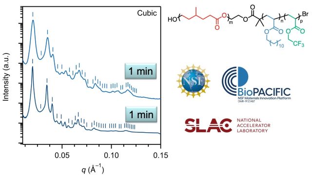
Above: ABC triblock terpolymer synthesized by ring-opening and atom transfer radical polymerization, comparing intensities as measured at the BioPACIFIC MIP and the SLAC National Accelerator Laboratory. Data courtesy of Elizabeth Murphy
Grazing Incidence SAXS (GISAXS)
Grazing Incidence SAXS (GISAXS) is a variation of traditional transmission SAXS, where the incoming X-ray radiation impinges on the sample at a grazing angle, typically close to the critical angle for total external reflection. The GI geometry results in a preferential illumination of only the top layer with a few nanometers thickness, thus enhancing surface scattering and making GISAXS particularly valuable for studying thin films and interfaces of nanostructured materials. Like most XRD setups, the GI technique is non-destructive and allows for in-situ studies, making it possible to monitor dynamic processes and changes in thin films and nanostructured materials over time. The grazing incidence mode can also be used in the WAXS mode (known as GIWAXS), which can be set automatically within the user interface. The high brilliance SAXS/WAXS platform instrument combines a high-brightness liquid metal-jet source, tunable beam-size, and a suite of in-house designed sample cells, enabling a multitude of experiments that previously required a synchrotron facility to be performed in the laboratory environment.

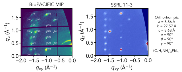
Above: A comparison of the 2D scattering pattern of a Perovskite crystal using the BioPACIFIC MIP SAXS/WAXS, and the same sample at a synchrotron source (SSRL). While the resolution and brilliance of the synchrotron image is expectedly better than the in-house image, all peaks present within the synchrotron exposure are also observed in the BioPACIFIC MIP diffractometer image (with exposure times on order of minutes), and both images give the same lattice parameters for the orthorhombic crystal.
