

Transmission Electron Microscopy Gains New Capabilities with Advanced Camera at CNSI
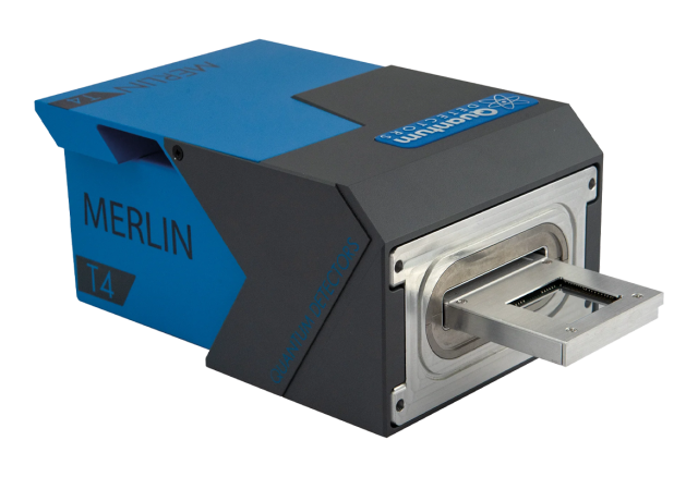
State-of-the-art Quantum Detectors Merlin T4 provides direct electron detection for finer control over data reconstruction and analysis
Investigators using transmission electron microscopes (TEM) to delve into nanoscale phenomena can run up against a number of obstacles. It can be difficult to capture data on ultrafast processes. Weak signals can be difficult to detect. And some samples are susceptible to electron-beam damage.
To overcome these challenges, the California NanoSystems Institute (CNSI) at UCLA has brought online the Quantum Detectors Merlin T4 hybrid pixel detector. The device offers advantages across numerous scientific applications — whether it’s mapping structures in new materials, characterizing features and defects of semiconductors or nanoparticles, or exploring beam-sensitive biomaterials, polymers, batteries or catalysts.
The Merlin T4 works in conjunction with the Thermo Fisher Scientific Spectra 300C at the National Science Foundation–funded BioPACIFIC Materials Innovation Platform. This addition extends the microscope’s capabilities for advanced diffraction, four-dimensional scanning TEM (4D-STEM), time-resolved measurements and low-dose characterization. Thanks to an event-based mode that records information about where and when each electron arrives with 200-picosecond precision, researchers gain finer control of how TEM data are reconstructed and analyzed over different time windows.
The Spectra 300C and Merlin T4 may be used by investigators on campus, from other universities or affiliated with industry.
“This new detector will significantly expand UCLA’s electron microscopy capabilities by combining fast, sensitive electron detection with exceptionally precise spatial and temporal measurements,” said Jianwei “John” Miao, a UCLA professor of physics and astronomy and a CNSI member. “It will enable advanced imaging and diffraction techniques, including electron ptychography, while creating new opportunities to resolve atomic arrangements and defects in quantum and energy materials and to develop low-dose cryogenic imaging of proteins and cellular structures. These capabilities could deepen our understanding of how structure and dynamics at the smallest scales govern material properties and biological function.”
Recent work, featured on the cover of Biophysical Journal, from scientists at CNSI used a similar camera at Lawrence Berkeley National Laboratory to study protein crystals, observing how they change due to electron irradiation. Experiments like this rely on the uniquely high framerates — exceeding 10,000 Hz — offered by these cameras.
The new camera opens up possibilities across multiple methods:
- For 4D-STEM, it creates information-rich datasets from two-dimensional diffraction patterns at each probe position.
- An acquisition rate that can exceed 40,000 effective frames per second enables electron diffraction measurements and applications requiring rapid sampling. The Merlin T4’s full-field readout and dual-counter pixel architecture reduces gaps during acquisition.
- With the ability to reconstruct sample phase from overlapping diffraction measurements, the instrument supports computational microscopy techniques such as ptychography.
- Direct, effectively noise-free electron counting and single-electron sensitivity expands the range of dose-sensitive specimens that can be mapped.
- Meanwhile, a conventional frame-based mode remains available when better-suited for an experiment.
The Merlin T4 software, developed in-house by the camera’s makers, is highly configurable. It supports real-time visualization, and can be integrated with Python-based analysis, EPICS and automation frameworks.
Potential users seeking consultation, proof-of-concept or information about how to access the Spectra 300C and Merlin T4 should reach out to biopacificmip@cnsi.ucla.edu.